

Signs and Solutions for Sleep Disorders
July 27, 2026
Creating a Personalized Health Plan for Preventive Cardiology
July 31, 2026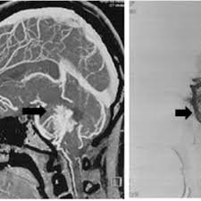
Neuroradiology studies the brain, spine, and nervous system through medical imaging, and it involves complex techniques. Specialists use these tools to spot problems, and they guide treatment with detailed pictures. When a cerebral aneurysm forms, imaging shows its size, shape, and location. Here is more information on neuroradiology techniques when they are used to assess cerebral aneurysms:
Cerebral Angiography Techniques
Cerebral angiography maps the blood vessels inside your brain. A doctor threads a thin catheter into an artery, and contrast dye highlights the vessels on X-ray. This neuroradiology method shows aneurysms with sharp detail. Since it offers precise views, teams use it before surgery.
Digital techniques build on this approach. They remove bone and tissue from the image, which provides a clear view of the vessels. The technique captures blood flow in real time, and physicians study each frame closely. Doctors use this data to measure an aneurysm accurately.
Angiography carries some risks that patients discuss with their care team. Bleeding at the catheter site happens occasionally, though teams monitor it carefully. Since the procedure is invasive, doctors weigh it against other options. Typical points doctors review include:
- Vessel narrowing or blockage
- Aneurysm neck width
- Arteries that are nearby
Computed Tomography Techniques
Computed tomography (CT) scans your head using rotating X-rays. A CT angiogram adds contrast dye, and the scanner produces layered images of the arteries. This test runs quickly in emergency settings. Doctors may order it first when they suspect a ruptured aneurysm.
CT angiography reconstructs vessels in three dimensions, and radiologists rotate the images on screen to view an aneurysm from every angle. Since the process takes minutes, teams gather answers fast during a crisis. The scan detects calcium and clotting within a vessel wall.
This tool provides detailed cross-sectional images of the body. The process is noninvasive and relies on advanced X-ray technology to visualize internal structures. While CT scans are quick and effective, they require careful interpretation by skilled radiologists to promote accurate diagnoses.
Magnetic Resonance Technology
Magnetic resonance imaging (MRI) uses strong magnets instead of radiation. The scanner maps soft tissue in detail, and it shows how blood moves through vessels. This method suits patients who avoid X-ray exposure. When time allows, doctors turn to MRI for a closer look.
MR angiography works without contrast dye in many cases. The scanner tracks moving blood and outlines each vessel clearly, and this technique reveals small aneurysms that other scans might miss. Since it avoids radiation, doctors sometimes choose it for repeat monitoring. Radiologists match the method to your specific situation, and factors that shape the choice include:
- Kidney function and dye tolerance
- Aneurysm size and location
- Urgency of the diagnosis
Visit a Neuroradiology Clinic Today
A neuroradiology clinic brings these tools together, and trained specialists read your images and explain the findings in plain terms. Early assessment gives your care team the details they need. Booking an appointment starts that process without delay. Talk with your doctor about which scan fits your needs; ask questions, review your options, and schedule a consultation with a neuroradiology team near you.